Medical Disclaimer
This page provides general health information for educational purposes and is not a substitute for professional medical advice, diagnosis, or treatment. Always consult a qualified healthcare provider for personal medical decisions. If you suspect a medical emergency, call your local emergency number immediately.
What is Lymphedema?
Lymphedema is a chronic condition characterized by abnormal swelling that occurs when the lymphatic system cannot properly drain lymph fluid from body tissues. It most commonly affects the arms or legs, but can also develop in the face, neck, chest, abdomen, or genitals.
The swelling happens when excess lymph fluid builds up in the interstitial spaces, causing tissue inflammation, discomfort, and—if left untreated—progressive skin and tissue changes.
Key Facts
- Lymphedema is typically progressive but can be managed effectively.
- Early detection improves outcomes and may prevent severe complications.
- There is no universal cure, but treatment can reduce symptoms significantly.
- Worldwide, it affects millions of people, including those treated for cancer and individuals with congenital lymphatic disorders.
Why the Lymphatic System Matters
A healthy lymphatic system continuously returns clear lymph fluid—containing proteins, fats, and immune cells—back into the bloodstream. When this drainage pathway is blocked or underdeveloped, fluid accumulates and lymphedema develops.
Left untreated, chronic fluid stagnation can lead to fibrosis, recurring infections, and reduced quality of life.
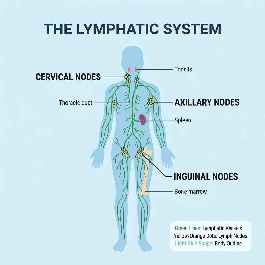
The Lymphatic System
The lymphatic system is a network of vessels, nodes, and organs that maintains fluid balance, absorbs dietary fats, and supports immune defense. It works alongside the circulatory system to remove waste and filter harmful substances.
How It Works
- Lymph vessels collect fluid from tissues and transport it toward the heart.
- Lymph nodes filter the fluid and trap bacteria, viruses, and abnormal cells.
- Lymphatic organs (spleen, thymus, tonsils) produce and mature immune cells.
- Muscle contractions and breathing help push lymph through the vessels.
When any part of this system is damaged, absent, or overwhelmed, lymphatic drainage slows and lymphedema can result.
Types of Lymphedema
Lymphedema is broadly classified as primary (caused by developmental abnormalities) or secondary (caused by damage to a previously normal lymphatic system).
| Feature | Primary Lymphedema | Secondary Lymphedema |
|---|---|---|
| Cause | Congenital or inherited malformation of lymph vessels/nodes | Damage to a healthy lymphatic system from surgery, radiation, trauma, infection, or cancer |
| Onset | May appear at birth, puberty, or later in life | Usually develops weeks, months, or years after the triggering event |
| Common Examples | Milroy disease, Meige disease, lymphedema tarda | Post-mastectomy lymphedema, post-radiation lymphedema, filariasis-related lymphedema |
| Prevalence | Rare; often runs in families | More common; the leading type worldwide |
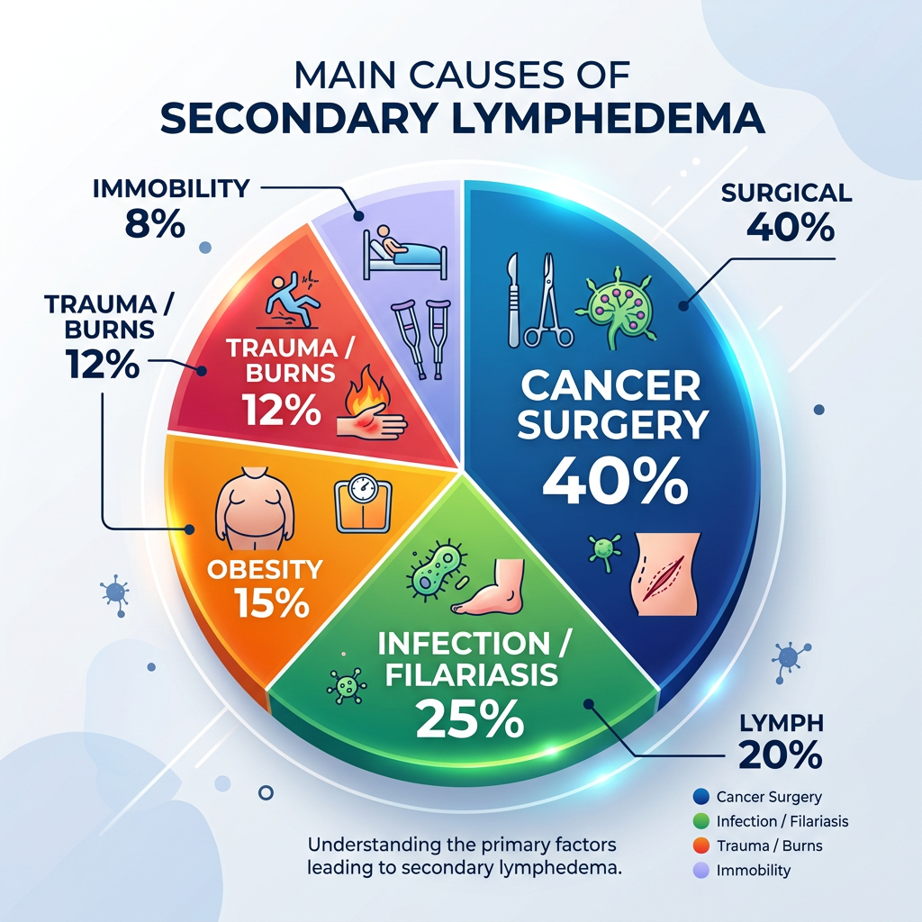
Main Causes of Lymphedema
Secondary lymphedema is most frequently caused by cancer treatment, infection, or physical trauma. Understanding the cause helps guide prevention and management strategies.
Cancer Surgery
Removal of lymph nodes during tumor surgery can interrupt lymphatic drainage.
Radiation Therapy
Radiation can scar and inflame lymphatic vessels and nodes, reducing flow.
Infection
Recurrent cellulitis or parasitic infections (e.g., lymphatic filariasis) can damage vessels.
Trauma & Injury
Burns, deep wounds, or fractures can disrupt local lymphatic pathways.
Obesity
Excess body weight increases lymphatic load and can impair drainage.
Venous Disease
Chronic venous insufficiency can overload the lymphatic system over time.
Lymphedema Stages
The International Society of Lymphology (ISL) classifies lymphedema into stages based on severity, tissue changes, and whether swelling is reversible.
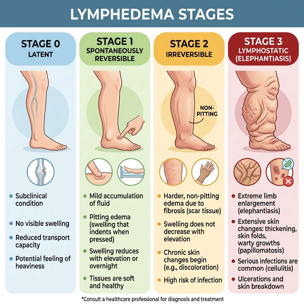
The lymphatic system is damaged but visible swelling is not yet present. Patients may experience heaviness, tightness, or fatigue in the limb. Early intervention during this stage can sometimes prevent progression.
Soft, pitting edema appears and often improves with elevation or overnight rest. Prompt treatment—compression, exercise, and manual lymph drainage—can produce excellent results at this stage.
Swelling does not resolve with elevation alone. Tissue becomes firmer (fibrotic) and pitting may lessen. Skin changes such as thickening or discoloration can begin. Ongoing therapy is needed.
Marked, non-pitting swelling with significant skin changes, folds, and fibrosis. The limb may become very large and heavy. Specialized care, including advanced compression and sometimes surgery, is required.
Normal Edema vs. Lymphedema
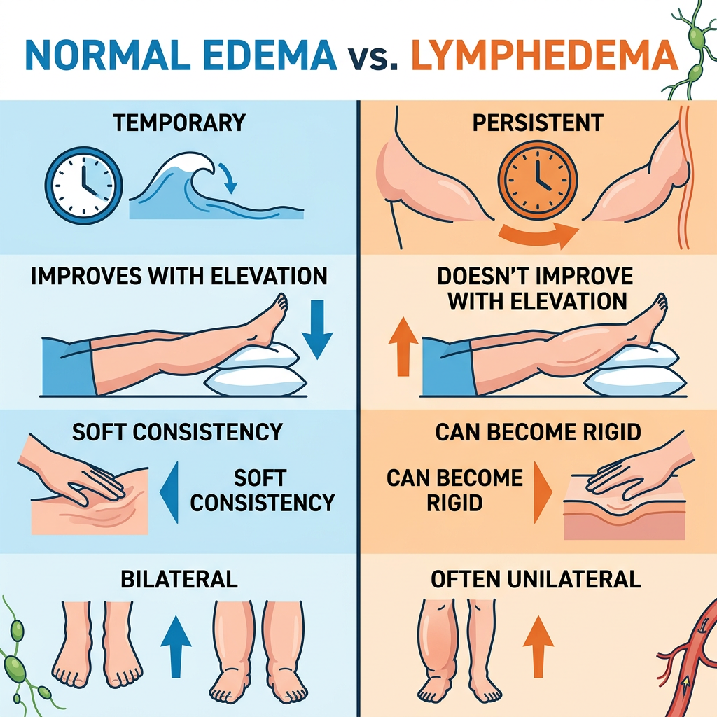
While both conditions involve swelling, they differ in cause, behavior, and treatment. Correct identification helps avoid delays in appropriate care.
| Characteristic | Normal Edema | Lymphedema |
|---|---|---|
| Typical cause | Salt intake, venous pressure, heart/kidney/liver issues | Lymphatic system damage, insufficiency, or obstruction |
| Pitting | Often pits when pressed | May pit early, but becomes non-pitting over time |
| Response to elevation | Usually improves quickly | May improve minimally or not at all in later stages |
| Skin changes | Usually minimal | Thickening, fibrosis, discoloration, and skin folds common |
| Infection risk | Generally lower | Higher risk of cellulitis and lymphangitis |
| Primary treatment | Treat underlying cause, diuretics, elevation | Complete Decongestive Therapy (CDT), compression, skin care |
Symptoms and Signs
Lymphedema symptoms can develop gradually. Recognizing them early allows for faster intervention and better long-term management.
Swelling
Persistent swelling in an arm, leg, hand, foot, or other body part that may worsen as the day progresses.
Heaviness or Tightness
A sensation of fullness, tight skin, or a heavy limb, often accompanied by reduced flexibility.
Restricted Movement
Difficulty bending or moving joints due to swelling and tissue stiffness.
Skin Changes
Thickening, hardening, discoloration, or development of skin folds and wart-like growths.
Recurrent Infections
Frequent episodes of cellulitis, lymphangitis, or skin infections in the affected area.
Aching or Discomfort
Dull aching, tingling, or discomfort in the affected limb or region.
Diagnosis & Stemmer's Sign
Diagnosis begins with a detailed medical history and physical examination. A healthcare provider will look for swelling patterns, skin changes, and risk factors such as cancer treatment or infection.
Common Diagnostic Steps
- Clinical history: Review of surgeries, radiation, infections, and family history.
- Physical exam: Measurement of limb circumference and assessment of skin texture.
- Imaging: Lymphoscintigraphy, ultrasound, MRI, or CT to evaluate lymphatic function.
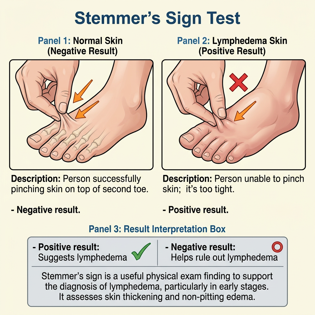
- Stemmer's sign: A simple clinical test used to distinguish lymphedema from other swelling.
About Stemmer's Sign
Stemmer's sign is tested by pinching the skin on the top of the foot or base of the fingers. In lymphedema, the thickened skin cannot be lifted or pinched into a fold. A positive sign strongly suggests lymphedema, while a negative sign does not rule out early disease.
Critical Alert Signs
Certain changes in the affected limb require prompt medical attention. Contact your healthcare provider if you notice any of the following:
Sudden Increase in Swelling
Rapid or noticeable swelling over hours or days without an obvious cause.
Redness or Warmth
New redness, warmth, or streaking on the skin can indicate infection.
Fever or Chills
Systemic symptoms alongside limb changes may signal cellulitis or sepsis.
Severe Pain
Significant or worsening pain that is not relieved by usual measures.
Open Wounds or Weeping
Skin breakdown, blisters, or leaking fluid increase infection risk.
Color Change
Blue, purple, or very pale skin, or coldness of the limb.
When to Seek Emergency Care
Some symptoms are medical emergencies. Call your local emergency number or go to the nearest emergency department immediately if you experience any of the following.
High Fever & Chills
Especially when combined with redness, warmth, or swelling of the affected limb.
Rapid Spreading Redness
Streaking red lines moving up the limb suggest a spreading infection.
Sudden Severe Pain
Acute, severe pain with swelling may indicate a blood clot or compartment syndrome.
Difficulty Breathing
Shortness of breath or chest pain requires immediate evaluation.
In the United States, dial 911. Use your local emergency number elsewhere.
Trusted Sources
- International Society of Lymphology. The diagnosis and treatment of peripheral lymphedema: 2020 Consensus Document of the International Society of Lymphology. Lymphology. 2020;53(3):100-115.
- National Lymphedema Network (NLN). Position Papers on Lymphedema Risk Reduction, Diagnosis, and Exercise. Available at: lymphnet.org.
- Mayo Clinic. Lymphedema: Symptoms and Causes. www.mayoclinic.org.
- Cancer.Net. Lymphedema: What People With Cancer Need to Know. American Society of Clinical Oncology. www.cancer.net.
- National Cancer Institute. Lymphedema (PDQ®)–Patient Version. www.cancer.gov.